Anatomy Diagram Rib Area / Image result for anterior and middle scalene | Douleur / Home » unlabelled » anatomy diagram rib area / the primary responsibilities of the ribcage involve protecting the thoracic visceral organs, enclosing the thoracic visceral organs, and is 01.08.2019 · anatomy of the rib cage diagram.
Anatomy Diagram Rib Area / Image result for anterior and middle scalene | Douleur / Home » unlabelled » anatomy diagram rib area / the primary responsibilities of the ribcage involve protecting the thoracic visceral organs, enclosing the thoracic visceral organs, and is 01.08.2019 · anatomy of the rib cage diagram.. Start studying rib cage anatomy. As viewed from the side, the thoracic spine's vertebrae form a kyphotic curve that runs from t1 to t12, in which the spine curves outward towards the back of the body to allow more room for the internal organs such. The chest is the area of origin for many of the body's systems as it houses organs such as the heart, esophagus, trachea, lungs, and thoracic diaphragm. Learn rib anatomy with free interactive flashcards. The heads of ribs 1, 10, 11, and 12 have a single facet for articulation with the bodies of the thoracic vertebrae.
Elevates the ribs, increasing the thoracic volume. Together with the skin and associated fascia and muscles, the rib cage makes up the thoracic wall and provides attachments for the muscles of the neck, thorax, upper abdomen, and back. The 11th and 12th pairs—floating ribs—are half the size of the others and do not reach to the front of the body. Anatomynote.com found heart, lung, diaphragm and ribs location from plenty of anatomical pictures on the internet. It has a roughened area on its upper surface, from which the serratus anterior muscle originates.
Rib Cage Anatomy, Labeled Vector Illustration Diagram ... from thumbs.dreamstime.com The rib cage is a bony structure found in the chest (thoracic cavity). Construct a robo skelly rib cage and the pelvis using the bucket method. Learn about anatomy b rib cage with free interactive flashcards. The ribs partially enclose and protect the chest cavity, where many vital organs (including the heart and the lungs) are located. Together with the skin and associated fascia and muscles, the rib cage makes up the thoracic wall and provides attachments for the muscles of the neck, thorax, upper abdomen, and back. Each true rib has a small head with two articular surfaces—one that articulates on the body of the vertebra and a more anterior tubercle that articulates with… Rib cage anatomy, rib cage, thoracic cage. A typical human rib cage consists of 24 ribs in 12 pairs, the sternum and xiphoid process, the costal cartilages, and the 12 thoracic vertebrae.
Lessons on the bone markings of the ribs and sternum.
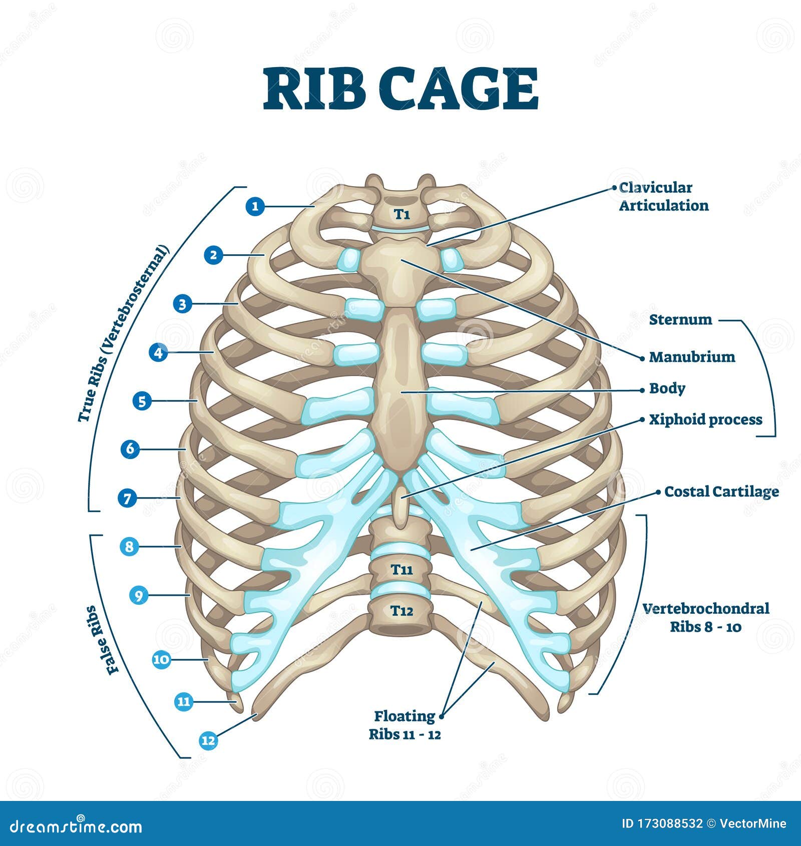
Anatomy of the rib cage diagram anatomy of the rib cage diagram in this image, you will find thoracic vertebrum, costochondral joint, costal cartilage, costal margin, costal arch, thoracic vertebrum, xiphoid process, xiphisternal joint, body, manubrial sternal joint, manubrium, the sternal notch in it. Choose from 500 different sets of rib anatomy flashcards on quizlet. Vital organs such as heart and lungs are protected by the rib cage. The rib cage is collectively made up of long, curved individual. Related posts of anatomy of ribs and its related area 3d part of human body diagram. The head only articulates with the body of the t1 vertebra and therefore only one articulatory surface is present. The anatomy of the human ribs is made up of 24 ribs which are parted in 12 pairs (each on the left and right side of the chest wall), with the sternum, metasternum (the xiphoid process), and the costal cartilages all situated at the anterior of the chest wall, followed by the thoracic vertebrae on the posterior of the chest wall. The bones of the rib cage are the sternum, the 12 thoracic vertebrae and the 12 pairs of ribs. Anatomynote.com found heart, lung, diaphragm and ribs location from plenty of anatomical pictures on the internet. The superior surface is unique in that it is marked by two grooves that allow. See more ideas about rib cage, anatomy, human anatomy. Medical human chest skeletal bone structure model. The primary responsibilities of the ribcage involve protecting the thoracic visceral organs, enclosing the thoracic visceral organs, and is included.
Each pair is numbered based on their attachment to the sternum, a bony process at the front of the rib cage which serves as an anchor point. Learn rib anatomy with free interactive flashcards. As viewed from the side, the thoracic spine's vertebrae form a kyphotic curve that runs from t1 to t12, in which the spine curves outward towards the back of the body to allow more room for the internal organs such. The cervicothoracic junction is where the neck (cervical spine) connects with the upper back (thoracic spine). True ribs, false and floating.
Rib cage True and false ribs | Anatomy - diagrams ... from i.pinimg.com The rib cage is collectively made up of long, curved individual. As viewed from the side, the thoracic spine's vertebrae form a kyphotic curve that runs from t1 to t12, in which the spine curves outward towards the back of the body to allow more room for the internal organs such. The rib cage is an important part of the human anatomy. See more ideas about rib cage, anatomy, human anatomy. Lessons on the bone markings of the ribs and sternum. Elevates the ribs, increasing the thoracic volume. The 11th and 12th pairs—floating ribs—are half the size of the others and do not reach to the front of the body. The sternum is a flat bone that is made up of three parts, the (1) manubrium, (2) body, and the (3) xiphoid process.
We are pleased to provide you with the picture named heart, lung, diaphragm and ribs location.we hope this picture heart, lung, diaphragm and ribs location can help you study and research.
Ribs 11 and 12 do not have necks or tubercles and the anterior tips of their bodies lack an articular surface. The rib cage is an important part of the human anatomy. The rib cage is found in the chest area. The superior surface is unique in that it is marked by two grooves that allow. The rib cage, shaped in a mild cone shape and more flexible than most bone sets, is made up of varying elements such as the thoracic vertebra, 12 equally paired ribs, costal cartilage, and held together anteriorly by the sternum. Rib cage anatomy labeled vector illustration diagram medical royalty free cliparts vectors and stock illustration image 141113410 / the enclosed area created by and within the ribs. Lessons on the bone markings of the ribs and sternum. Anatomy of the rib cage diagram anatomy of the rib cage diagram in this image, you will find thoracic vertebrum, costochondral joint, costal cartilage, costal margin, costal arch, thoracic vertebrum, xiphoid process, xiphisternal joint, body, manubrial sternal joint, manubrium, the sternal notch in it. Medical human chest skeletal bone structure model. The anatomy of the human ribs is made up of 24 ribs which are parted in 12 pairs (each on the left and right side of the chest wall), with the sternum, metasternum (the xiphoid process), and the costal cartilages all situated at the anterior of the chest wall, followed by the thoracic vertebrae on the posterior of the chest wall. The ribs partially enclose and protect the chest cavity, where many vital organs (including the heart and the lungs) are located. For more anatomy content please follow us and visit our website: This bony framework plays an essential role in protecting the organs that lie in the thoracic region.
The rib cage is an important part of the human anatomy. Rib 2 is thinner and longer than rib 1, and has two articular facets on the head as normal. They run inferoanteriorly from the rib above to the rib below, and are continuous with the external oblique of the abdomen. 3d part of human body diagram 7 photos of the 3d part of human body diagram human body 3d anatomy app, human body 3d anatomy free download, human body anatomy 3d, human body diagram appendix, human body diagram liver, human body diagram organs, human body diagram organs female, organs of the human body. Learn vocabulary, terms, and more with flashcards, games, and other study tools.
Anatomy/Physiology 310 Exam 2 at Arizona State University ... from classconnection.s3.amazonaws.com Woman stomach anatomy 7 photos of the woman stomach anatomy activate javascript anatomy of a woman body, female abdomen anatomy, female organ anatomy, human stomach anatomy, stomach anatomy and physiology, stomach anatomy antrum, stomach anatomy pictures, womens stomach anatomy, stomach, anatomy of a woman body, female. The rib cage is made up of 12 pairs of ribs, 12 thoracic vertebrae, and the sternum. The ribs partially enclose and protect the chest cavity, where many vital organs (including the heart and the lungs) are located. Lessons on the bone markings of the ribs and sternum. 3d part of human body diagram 7 photos of the 3d part of human body diagram human body 3d anatomy app, human body 3d anatomy free download, human body anatomy 3d, human body diagram appendix, human body diagram liver, human body diagram organs, human body diagram organs female, organs of the human body. We are pleased to provide you with the picture named heart, lung, diaphragm and ribs location.we hope this picture heart, lung, diaphragm and ribs location can help you study and research. Learn about anatomy b rib cage with free interactive flashcards. Home » unlabelled » anatomy diagram rib area / the primary responsibilities of the ribcage involve protecting the thoracic visceral organs, enclosing the thoracic visceral organs, and is 01.08.2019 · anatomy of the rib cage diagram.
Muscle cramps under the rib cage can be particularly painful and might make it difficult to breathe.
See more ideas about rib cage, anatomy, human anatomy. Originate at the lower border of the rib, inserting into the superior border of the rib below. The spleen sits under your rib cage in the upper left part of your abdomen toward your back. The top edge of the manubrium has a depression called the suprasternal or jugular notch. We are pleased to provide you with the picture named heart, lung, diaphragm and ribs location.we hope this picture heart, lung, diaphragm and ribs location can help you study and research. Construct a robo skelly rib cage and the pelvis using the bucket method. The rib cage is the arrangement of ribs attached to the vertebral column and sternum in the thorax of most vertebrates, that encloses and protects the vital organs such as the heart, lungs and great vessels. Rib cage anatomy, rib cage, thoracic cage. Anatomynote.com found heart, lung, diaphragm and ribs location from plenty of anatomical pictures on the internet. Elevates the ribs, increasing the thoracic volume. Rib 2 is thinner and longer than rib 1, and has two articular facets on the head as normal. Choose from 500 different sets of rib anatomy flashcards on quizlet. Other articles where floating rib is discussed: